Some uses of radioactivity: in medicine
Sterilisation surgical instruments
Gamma rays are high energy electromagnetic waveA transverse wave caused by oscillations in an electromagnetic field. which are only stopped by thick lead.
This means they can easily pass through medical equipment, such as syringes.
As gamma rays pass through the packaging and syringe, they will kill viruses and bacteria which contaminate the syringe.
As long as the equipment remains in a sealed plastic pack it will remain free of viruses and bacteria and be safe for use with patients.
The gamma ray source used should have a long half-lifeThe half-life of a radioactive source is the time taken for its activity to fall to half of the original activity. Half-life is measured in seconds. so that the hospital does not have to replace it too frequently
Advantages
- sterilisation can be done without high temperatures;
- it can be used to kill bacteria on things that would melt e.g. plastic syringes.
Disadvantages
- it may not kill all bacteria on an object;
- it can be very harmful - standing in the environment where objects are being treated by radiation could expose peopleÔÇÖs cells to damage and possibly cancer.
Radiotherapy: killing cancerous tumours

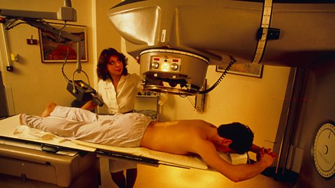
Although ionising radiationRadiation that is able to remove electrons from atoms or molecules to produce positively charged particles called ions. can cause cancer, high doses can be directed at cancerous cells to kill them.
This is called radiotherapy.
About 40 per cent of people with cancer undergo radiotherapy as part of their treatment.
It is administered in two main ways:
- from outside the body using X-rays or gamma rays from radioactive cobalt;
- from inside the body by putting radioactive materials into the tumour, or close to it.
Gamma knife
- beams of gamma rays, called a gamma knife, can be used to kill the cancerous tumour deep inside the body;
- these beams are aimed at the tumour from many different directions to maximise the dose on the tumour but to minimise the dose on the surrounding soft tissue. This technique can damage healthy tissue, so careful calculations are done to establish the best dose - enough to kill the tumour, but not so much so that the healthy tissue is damaged.
The gamma ray source used should have a long half-life so that the hospital does not have to replace it too frequently
Radioactive Tracers
In some cases, injected radioactive sources (such as technetium-99) can be used as tracers to make soft tissues, such as blood vessels or the kidneys, show up through medical imaging processes.
Iodine-131 is used as a radioactive tracer to investigate the thyroid gland.
The radioactive source emits gamma rays that easily pass through the body to a detector outside the body, for example a ÔÇÿgamma cameraÔÇÖ.
In this way, the radioactive source can be followed as it flows through a particular organ in the body.
Changes in the amount of gamma rays emitted from different parts would indicate how well the isotope is flowing, or if there is a blockage.
The source used must:
- be a source of gamma rays so that they pass out through the body to be detected by the gamma camera or GM tube;
- have very short half-lives - sources used typically have half-lives of hours so after a couple of days there will hardly be any radioactive material left in a personÔÇÖs body;
- not be poisonous.